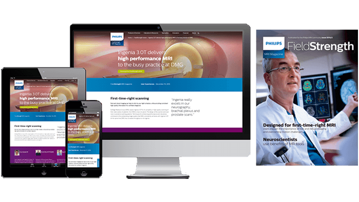
FieldStrength MRI magazine
User experiences - December 19, 2015
Fast and robust imaging can help to stick to your tight schedule, while providing consistent high quality information for confident diagnosis.
DuPage Medical Group (DMG) values Ingenia 3.0T for its versatility in high quality scanning of their wide variety of patients, from MSK to neuro to pelvis. The design of the scanner and scan room is aimed at soothing patients. Techniques for enhancing performance and robustness contribute to the outstanding image quality that DMG consistently achieves with Ingenia 3.0T. At the same time DMG has increased throughput on its Ingenia.
“Ingenia really excels in our neurography, brachial plexus and prostate scans.”
Yazan has been a radiologist for 16 years. He was educated at St. Louis University School of Medicine and performed his residency at Louisiana State University School of Medicine, with fellowships at University of Virginia School of Medicine and University of Washington School of Medicine.
Received his BS from Southern Illinois University at Carbondale. Upon graduation he completed an internship and three years of employment at Northwestern Memorial Hospital. He has been with DuPage Medical Group for twelve years and is now the Lead MRI Technologist, where he oversees five magnets with 18 MRI technologists.
Received his AS in Radiography from Ferris State University. He has worked with MRI since graduation, beginning his career at KNI/Southwest Imaging Michigan and was a dedicated Philips MRI user. Prior to joining DuPage Medical Group in 2006, he worked at an outpatient facility in North Carolina.
Ingenia helps surmount challenges of a busy practice
DuPage Medical Group (Downers Grove, Illinois, USA) is a physician-owned group outside Chicago that performs about 2,000 MRI exams per month. The radiology department in Lisle, Illinois is using a Philips Ingenia 3.0T in addition to MRI scanners from other vendors.
“We have to scan a wide range of patients and we want to emphasize both image quality and speed. Taking into account these challenges we also need to maintain our time slots at 30 minutes. For routine MR, we maximize acquisition speed for workflow efficiency,” says Yazan Kaakaji, MD, radiologist at DMG. “Ingenia 3.0T is a comprehensive scanner that performs according to these needs.”
“Patient experience is another important factor. We don’t have a sedation policy, so we wanted a fast scanner and a soothing experience for our patients, particularly for those who are relatively claustrophobic. Ingenia 3.0T with the wide bore and Ambient cove lighting provides the patient experience that we desired. We get positive comments from patients on it.”
Image quality is “excellent,” “phenomenal,” “stellar”
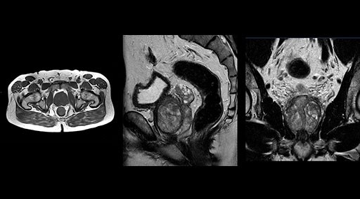
Patrick Duffy BS, RT (R) MR is Lead Technologist at DMG. “We are getting phenomenal image quality on all types of exams,” he says. “Our MSK is stellar, and so is our abdominal work. Ingenia excels at feet, hands and fingers. We do enterographies with great results. With the combination of the 3.0T magnet and the digital coils, we are able to scan prostates without an endorectal coil while still obtaining high quality results. This is a comforting experience for our male patients. We scan many obese patients, and the Ingenia does a tremendous job because of MultiTransmit, which reduces dielectric shading for more confident diagnosis. Our technologists really enjoy scanning on the Ingenia. We also have ordering physicians who specifically want their patients scanned on the Ingenia because of the results of our imaging.
“Obviously, the diagnostic capability is most important,” says Dr. Kaakaji. “Ingenia’s image quality is excellent and in follow-up studies, Ingenia provides good consistency between scans.
“Without using an endorectal coil we do our prostate MR at 0.5 mm resolution, following the European society of urology protocol [1]. For certain joints we use a virtual arthroscopy protocol with 1 mm pixel size and 2 mm slice thickness. Ingenia really excels in our neurography, brachial plexus and prostate scans. Our neurologists insist on using our 3.0T for those,” Dr. Kaakaji adds.
“The image quality is phenomenal. Robust, clear, homogenous, not obscured by dielectric shading,” adds technologist Ryan Sybesma, RT (R) MR. “Ingenia is a high performance workhorse.”
“We’ve added two daily slots to the schedule after experiencing Ingenia’s performance.”
Clinical cases
“With mDIXON TSE we get good results the first time – that’s significant, because repeating one scan could put us behind schedule.”
mDIXON TSE fat suppression helps DMG reduce repeats and supports diagnostic confidence
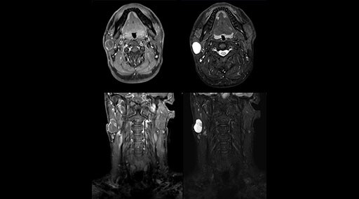
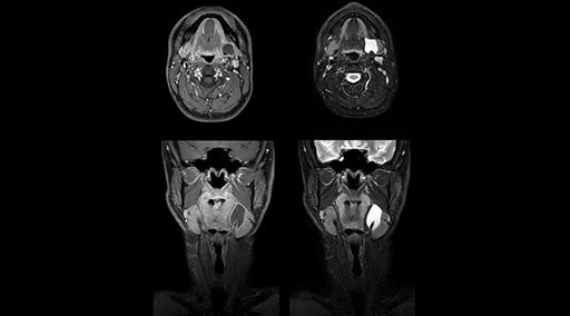
“Our DMG Lisle location includes a cancer center, so soft tissue neck scans, brachial plexus scans, and prostate scans are common. For these exams, mDIXON TSE provides excellent images with and without fat suppression all while helping us reduce repeats and work more efficiently,” Mr. Duffy says.
“With the 2-echo Philips mDIXON TSE the timing is short and the fatsat is very robust. The biggest thing is that you know your fat suppression will be good, even in thin patients or large patients that are off-center,” Mr. Sybesma says.
“Since we work in fixed time slots, not having to repeat scans is key for us,” Mr. Duffy adds. “With mDIXON TSE we get high quality results the first time – unless of course the patient absolutely jumps off the table. For us, that’s significant, because just a single repeat scan could put us behind schedule.
“mDIXON TSE raises our diagnostic confidence with its homogeneous
fat suppression. Neck exams and rheumatology patients are two examples where mDIXON TSE is especially useful,” Dr. Kaakaji says. “For us it’s also an efficiency boost in exams where we need pre and post T1-weighted images with great fat suppression.”
MultiVane XD helps eliminate motion for first-time-right scanning
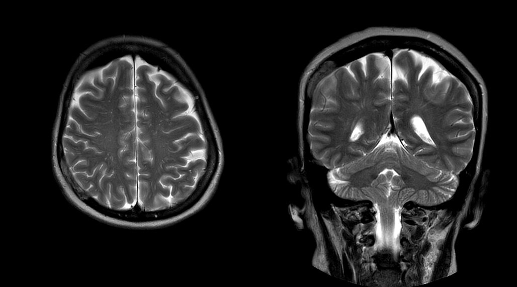
DMG recognizes MultiVane XD motion compensation is another Philips technique that contributes to image quality and scan efficiency. “We run MultiVane XD for motion-free imaging on almost all our T2-weighted brain scans, just to reduce any repeats we might get. We know our non-contrast brain scans are going to take 20 minutes almost every time,” Mr. Duffy says.
“Using MultiVane XD still allows us to turn on dS SENSE, which significantly cuts scan time compared to what we were doing before,” he adds. “We went from a 2.5 or 3-minute scan to a 1.5-minute scan with no loss in image quality. So, it not only reduces the motion, but also reduces scan time. That gives us a little bit of extra time to speak to our patients and explain the exam a little more.”

Workflow benefits help increase throughput
The workflow benefits of Ingenia 3.0T are indisputable. “We especially like it in spine,” says Dr. Kaakaji. “We have a 12-minute spine protocol for non-contrast lumbar and cervical. We perform our lumbar spines feet first, just using the posterior coil integrated in the table. With the tiltable head coil Ingenia makes it easy to image elderly or kyphotic patients because we can raise their head. We can do spines in about 10 minutes if we have to, and there’s no contrast involved.”
“I think it’s also a comfortable scanner for patients – they are in there for sometimes less than 15 minutes. That absolutely helps with throughput. We’ve added two daily slots to the schedule after experiencing Ingenia’s performance.”
Techs like working with Ingenia
“The technologists enjoy Ingenia’s user interface and easy scanning,” says Mr. Duffy. “I have techs at different levels who all do a great job on Ingenia. I think it’s an easy scanner to learn, yet at the same time it’s one of the most advanced scanners.
Mr. Sybesma agrees. “Ingenia is so versatile. I love the integrated Posterior coil in the table; the long field of view is really helpful, and we can help patients by having them go in feet first instead of head first. The coils are easy to use, and just give us phenomenal images. SmartAssist’s automatic coil elements selection is excellent – there’s no running a 5-minute scan with the wrong coil selected – it definitely helps a lot. Radiologists and referring physicians compliment us all the time on our phenomenal knee images.”
“Ingenia is also my preferred scanner to do breast imaging. Using the breast coil, we haven’t had inhomogeneity issues at all. We also use SmartExam for our breast patients, for consistent, reproducible scans. Our breast images look just remarkable,” he adds.
Patients appreciate Ingenia design
“Of course, patient satisfaction is vital to a successful practice,” Mr. Duffy says. “I believe the Ingenia looks very soothing to most people. I get comments from patients as soon as they enter the magnet room. Compliments range anywhere from the aesthetics of the wide bore scanner to how cool the room looks with the special LED cove lighting. I believe it really puts patients at ease, especially if they’re apprehensive of the scan to start with.”
Summarizing how Ingenia excels at DMG
“To me the top three strengths of Ingenia are its versatility to do exams without issues, the phenomenal image quality and also the support from Philips,” says Mr. Sybesma.
“When recommending Ingenia to other users, I would highlight the
image quality, the consistency of all exams and the overall appearance of the machine,” says Mr. Duffy. “Ingenia is an excellent MRI scanner that will perform to the level of every user – from basic MSK to advanced scanning. It’s easy to run and produces quality images. We run a tight ship, and that’s all we can ask for.”
Dr. Kakaaji feels well equipped for the future with Ingenia 3.0T. “In our institution we see MR utilization expanding at the expense of other diagnostic imaging and within MR we see body and oncology applications expanding. I think Ingenia 3.0T is tailored to both. We are ready to move forward.”
Reference
1. Barentsz JO, Richenberg J, Clements R, Choyke P, Verma S, Villeirs G, Rouviere O, Logager V, Fütterer. JJ ESUR prostate MR guidelines 2012. Eur Radiol (2012) 22:746–757
Results from case studies are not predictive of results in other cases. Results in other cases may vary. Results obtained by this facility may not be typical for all facilities. Images that are not part of User experiences articles and that are not labeled otherwise are created by Philips.
“mDIXON TSE raises our diagnostic confidence with its homogeneous fat suppression”
