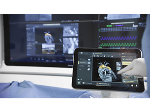
VesselNavigator

Reduce your need for contrast medium in endovascular procedures

Encontrar produtos semelhantes

VesselNavigator allows reuse of 3D vascular anatomical information from existing CTA and MRA datasets as a 3D roadmap overlay on a live X-ray image. With its excellent visualization, VesselNavigator provides an intuitive and continuous 3D roadmap to guide you through vasculature during the entire procedure.

Características
3D image fusion || A

3D image fusion in endovascular procedures

Studies¹,² have shown the advantages of a 3D perspective for catheter and device guidance. With VesselNavigator, clinicians can easily segment 3D vessel structures from existing CT/MR datasets and fuse them with live X-ray for guidance.

3D image fusion in endovascular procedures

Studies¹,² have shown the advantages of a 3D perspective for catheter and device guidance. With VesselNavigator, clinicians can easily segment 3D vessel structures from existing CT/MR datasets and fuse them with live X-ray for guidance.

3D image fusion in endovascular procedures

Studies¹,² have shown the advantages of a 3D perspective for catheter and device guidance. With VesselNavigator, clinicians can easily segment 3D vessel structures from existing CT/MR datasets and fuse them with live X-ray for guidance.
Reduction in contrast usage || B

Reduction in contrast usage

Average contrast medium usage was reduced by 72% in a study¹ that used Philips CTA image fusion guidance during endovascular repair of complex aortic aneurysms. No intraprocedural contrast agent injection was required to create a roadmap.

Reduction in contrast usage

Average contrast medium usage was reduced by 72% in a study¹ that used Philips CTA image fusion guidance during endovascular repair of complex aortic aneurysms. No intraprocedural contrast agent injection was required to create a roadmap.

Reduction in contrast usage

Average contrast medium usage was reduced by 72% in a study¹ that used Philips CTA image fusion guidance during endovascular repair of complex aortic aneurysms. No intraprocedural contrast agent injection was required to create a roadmap.
Potential to reduce procedure time || C

Potential to reduce procedure time

Philips CTA Image Fusion Guidance may lead to shorter procedure times. A study of 62 patients² showed an average reduction in procedure time from 6.3 to 5.2 hours during FEVAR/BEVAR procedures using Philips CTA image fusion guidance.

Potential to reduce procedure time

Philips CTA Image Fusion Guidance may lead to shorter procedure times. A study of 62 patients² showed an average reduction in procedure time from 6.3 to 5.2 hours during FEVAR/BEVAR procedures using Philips CTA image fusion guidance.

Potential to reduce procedure time

Philips CTA Image Fusion Guidance may lead to shorter procedure times. A study of 62 patients² showed an average reduction in procedure time from 6.3 to 5.2 hours during FEVAR/BEVAR procedures using Philips CTA image fusion guidance.
Moves in sync to new positions || D

Moves in sync to new positions

During the procedure, VesselNavigator provides a real-time overlay that moves in sync with any projection, table and system position. This can reduce the need to make additional contrast enhanced runs to create new roadmaps.

Moves in sync to new positions

During the procedure, VesselNavigator provides a real-time overlay that moves in sync with any projection, table and system position. This can reduce the need to make additional contrast enhanced runs to create new roadmaps.

Moves in sync to new positions

During the procedure, VesselNavigator provides a real-time overlay that moves in sync with any projection, table and system position. This can reduce the need to make additional contrast enhanced runs to create new roadmaps.
Superb image quality || D

Superb image quality

Create overlays based on a variety of superb volume rendering visualization options. These can be customized according to user preferences.

Superb image quality

Create overlays based on a variety of superb volume rendering visualization options. These can be customized according to user preferences.

Superb image quality

Create overlays based on a variety of superb volume rendering visualization options. These can be customized according to user preferences.
Anatomical || D

Anatomical ring markers

Ring markers can be added to the segmented image to indicate the ostia and landing zone and to define planning angles. These markers are visualized during the procedure for guidance.

Anatomical ring markers

Ring markers can be added to the segmented image to indicate the ostia and landing zone and to define planning angles. These markers are visualized during the procedure for guidance.

Anatomical ring markers

Ring markers can be added to the segmented image to indicate the ostia and landing zone and to define planning angles. These markers are visualized during the procedure for guidance.
  • 3D image fusion || A
  • Reduction in contrast usage || B
  • Potential to reduce procedure time || C
  • Moves in sync to new positions || D
Veja todos os recursos
3D image fusion || A

3D image fusion in endovascular procedures

Studies¹,² have shown the advantages of a 3D perspective for catheter and device guidance. With VesselNavigator, clinicians can easily segment 3D vessel structures from existing CT/MR datasets and fuse them with live X-ray for guidance.

3D image fusion in endovascular procedures

Studies¹,² have shown the advantages of a 3D perspective for catheter and device guidance. With VesselNavigator, clinicians can easily segment 3D vessel structures from existing CT/MR datasets and fuse them with live X-ray for guidance.

3D image fusion in endovascular procedures

Studies¹,² have shown the advantages of a 3D perspective for catheter and device guidance. With VesselNavigator, clinicians can easily segment 3D vessel structures from existing CT/MR datasets and fuse them with live X-ray for guidance.
Reduction in contrast usage || B

Reduction in contrast usage

Average contrast medium usage was reduced by 72% in a study¹ that used Philips CTA image fusion guidance during endovascular repair of complex aortic aneurysms. No intraprocedural contrast agent injection was required to create a roadmap.

Reduction in contrast usage

Average contrast medium usage was reduced by 72% in a study¹ that used Philips CTA image fusion guidance during endovascular repair of complex aortic aneurysms. No intraprocedural contrast agent injection was required to create a roadmap.

Reduction in contrast usage

Average contrast medium usage was reduced by 72% in a study¹ that used Philips CTA image fusion guidance during endovascular repair of complex aortic aneurysms. No intraprocedural contrast agent injection was required to create a roadmap.
Potential to reduce procedure time || C

Potential to reduce procedure time

Philips CTA Image Fusion Guidance may lead to shorter procedure times. A study of 62 patients² showed an average reduction in procedure time from 6.3 to 5.2 hours during FEVAR/BEVAR procedures using Philips CTA image fusion guidance.

Potential to reduce procedure time

Philips CTA Image Fusion Guidance may lead to shorter procedure times. A study of 62 patients² showed an average reduction in procedure time from 6.3 to 5.2 hours during FEVAR/BEVAR procedures using Philips CTA image fusion guidance.

Potential to reduce procedure time

Philips CTA Image Fusion Guidance may lead to shorter procedure times. A study of 62 patients² showed an average reduction in procedure time from 6.3 to 5.2 hours during FEVAR/BEVAR procedures using Philips CTA image fusion guidance.
Moves in sync to new positions || D

Moves in sync to new positions

During the procedure, VesselNavigator provides a real-time overlay that moves in sync with any projection, table and system position. This can reduce the need to make additional contrast enhanced runs to create new roadmaps.

Moves in sync to new positions

During the procedure, VesselNavigator provides a real-time overlay that moves in sync with any projection, table and system position. This can reduce the need to make additional contrast enhanced runs to create new roadmaps.

Moves in sync to new positions

During the procedure, VesselNavigator provides a real-time overlay that moves in sync with any projection, table and system position. This can reduce the need to make additional contrast enhanced runs to create new roadmaps.
Superb image quality || D

Superb image quality

Create overlays based on a variety of superb volume rendering visualization options. These can be customized according to user preferences.

Superb image quality

Create overlays based on a variety of superb volume rendering visualization options. These can be customized according to user preferences.

Superb image quality

Create overlays based on a variety of superb volume rendering visualization options. These can be customized according to user preferences.
Anatomical || D

Anatomical ring markers

Ring markers can be added to the segmented image to indicate the ostia and landing zone and to define planning angles. These markers are visualized during the procedure for guidance.

Anatomical ring markers

Ring markers can be added to the segmented image to indicate the ostia and landing zone and to define planning angles. These markers are visualized during the procedure for guidance.

Anatomical ring markers

Ring markers can be added to the segmented image to indicate the ostia and landing zone and to define planning angles. These markers are visualized during the procedure for guidance.

You are about to visit a Philips global content page

Continue

You are about to visit a Philips global content page

Continue

Nosso site pode ser melhor visualizado com a versão mais recente do Microsoft Edge, Google Chrome ou Firefox.

É profissional de saúde?
Por favor, selecione a opção

Aviso:

A informação incluída nesta página destina-se exclusivamente a profissionais de saúde.